Research frontiers | Quantitative analysis of glutathione and its associated spontaneous neuronal activity in depressive disorders and compulsive disorders
Abstract
Glutathione (GSH) and glutamate (Glu) in the medial prefrontal cortex (mPFC) and precuneate were quantified by using a combination of magnetic resonance spectroscopy (MRS) and resting state functional magnetic resonance imaging using MEGA PRESS, and healthy controls (HCs, N=30), depressive disorders (MDD, MDD) were investigated. Relationship between glutathione levels and intrinsic neuronal activity and clinical symptoms in the three groups (N=28) and OCD (N=28).
Compared with HCs, the mPFC and precuneiform glutathione concentrations were lower in both MDD and OCD groups.
In healthy people, glutathione is positively correlated with glutamate levels and fractional amplitude of low-frequency fluctuations (fALFF) in both regions.
A weak positive association was shown between glutamate and fALFF values in both groups of patients. Glutathione levels were negatively associated with depressive symptoms and obsessive-compulsive symptoms in MDD and OCD patients, respectively.
These results suggest that the reduction of glutathione levels and the imbalance between glutathione and glutamate may increase oxidative stress and alter neurotransmitter signal transduction, leading to the breakdown of glutathione-related neurochemist-neuron coupling, mediating the occurrence and development of MDD and OCD diseases.
INTRODUCTION
Due to the imbalance between the production and degradation of reactive oxygen species, excess reactive oxygen species can lead to oxidative stress.
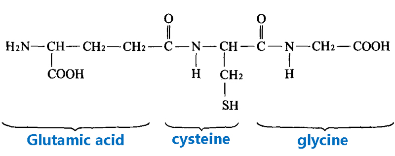
The most important and abundant antioxidant is glutathione, which protects against a variety of oxidative damage.
In the brain, neurons require high levels of glutathione to resist oxidative stress and maintain optimal brain function.
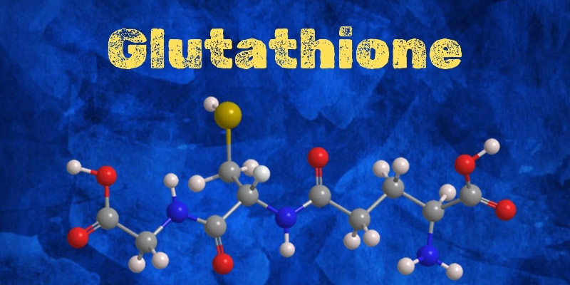
With the further acceptance of oxidative stress theory, people began to pay attention to the role of brain glutathione level in the occurrence and development of mental illness.
In studies using magnetic resonance spectroscopy (MRS) to investigate cortical glutathione levels in patients with depressive disorder, the results have been inconsistent, with some studies showing that the level of glutathione in patients with depressive disorder is lower than that in healthy people, and is associated with anhedonia, which is recovered after antidepressant treatment;
Other studies have found higher or no difference in glutathione levels.
As for glutathione levels in the cortex of people with compulsive disorder, only one study found that glutathione /Cr levels in the cingulate cortex after compulsive disorder were significantly lower than in healthy people.
Overall, the main reason for the rarity and inconsistency is the lack of accurate measurement methods.
Measuring glutathione using MRS Remains challenging due to the low concentration of glutathione in the brain (1.5-3 mmol/L), low signal-to-noise ratio, and severe spectral overlap between metabolites of different peak intensivities.
Many methods have been proposed to assess glutathione concentration in the human brain. For example, one of the latest MRS Acquisition sequences, MEGA-PRESS, uses subtraction of two repeated measurements to enhance the glutathione signal and is more sensitive than the PRESS method. This technique is also considered a standard for sequencing MRS Of other metabolites such as glutamic acid and gamma-aminobutyric acid.
To our knowledge, there was a recent study that used MEGA-PRESS to detect GSH levels for MDD, but not for OCD.
In the human brain, the relationship between glutathione and neurophysiology remains underexplored, not only in depressive disorders and compulsive disorders, but also in other neuropsychiatric disorders.
In addition to functional connectivity (FC), resting state functional magnetic resonance imaging (rs-fMRI) also provides low-frequency wave amplitude (ALFF) information, which measures the baseline strength of low-frequency oscillations of blood oxygen-level dependent signals that can reflect regional characteristics of intrinsic neural activity.
Variations in ALFF and fractional low frequency amplitude (fALFF) values in MDD and OCD have been the subject of several recent rs-fMRI studies.
Changes in ALFF and fALFF in two diseases have been reported in different brain regions.
Since neurotransmitters affect the activity of neurons, forming microscopic neural circuits, the connectivity of the brain represents the state of the macro-scale neural network.
To gain a comprehensive and in-depth understanding of brain function, it is necessary to consider the interaction between neurotransmitter concentrations (i.e., glutathione, Glu in this study) and local brain activity.

In this study, MEGA-PRESS MRS Was combined with rs-fMRI.
(1) Quantified glutathione and Glu concentrations in mPFC and precuneus;
(2) To study the relationship between brain glutathione and Glu;
(3) To investigate the relationship between glutathione level and intrinsic neuronal activity and clinical symptoms in HCs, MDD and OCD groups.
methodology
1. Scale:
- PHQ-9 (Patient Health Questionnaire-9)
- GAD-7 (Generalized Anxiety Disorder Scale -7 items)
- CES-D (Self-Rating Scale for Depression by Flow Center)
- Y-BOCS (Yale-Brown Obsessive Symptom Scale, Self-reported version)
- OCI-R (Compulsive Scale – Revised Edition)
2. Magnetic resonance parameters and sequences:
(1) Instrument: 3TGE SIGNA Architect scanner, 48 channel head coil
(2) Anatomical images: 3DT1-weighted images
(3) Function image:
Sequence: T2-weighted gradient echo planar imaging
Repetition time (TR) : 2,000 milliseconds
Echo time (TE) : 30 ms
Time series: 240
(4) Spectral sequence: MEGA-PRESS
Repetition time (TR) : 2,000 milliseconds
Echo time (TE) : 79 ms
Voxel size: 2×2×2 cm³
Location: mPFC (medial prefrontal cortex) and precuneus
(5) Correction of cerebrospinal fluid composition
3. MRS Data Processing:
After window processing and frequency and phase correction of spectral data, LCModel was used for spectral fitting.
Metabolite concentrations were calculated by normalizing and setting reference values, and data quality was assessed by signal-to-noise ratio and Crnar-RAO lower limits.
4. re-fMRI data processing:
The rest state functional images were preprocessed by SPM and the computational anatomy toolbox, including slice timing correction, realalignment, coregistration, tissue segmentation, standardization, and smoothing.
fALFF analysis was performed using the CONN toolbox, with frequencies ranging from 0.01 to 0.1 Hz, and the effects of white matter, cerebrospinal fluid, and motion effects were removed during the denoising step.

Monomeric elements of the mPFC and precuneus were normalized to MNI (Montreal Neurological Institute, a brain imaging coordinate system) to create regions of interest.
5. Data analysis:
SPSS 25 was used for statistical analysis.
One-way analysis of variance (ANOVA) was used to compare demographic characteristics, psychological status, fALFF, glutathione and Glu concentrations between groups.
Pearson correlation analysis examined the relationship between glutathione, Glu, fALFF, and psychological symptoms in each group, using the GAD-7 score as a covariate in the MDD group and the PHQ-9 score as a covariate in the OCD group.
All results were adjusted for p values using the Benjamini-Hochberg procedure.
result
1. Sample characteristics
No significant group differences were found between the groups in terms of age, sex and educational attainment.
Postmortem analysis showed that MDD group and OCD group had higher scores than HC group.
The PHQ-9 score in MDD group was higher than that in OCD group (P=0.017), and the Y-BOCS score in OCD group was higher than that in MDD group (P<0.001).

2. Quantitative results of glutathione and glutamate
Glutathione concentration (CRLB) in mPFC of HC, MDD and OCD groups was 1.36±0.25 (8.1%), 1.19±0.21(8.5%) and 1.14±0.31 (8.4%), respectively. Precuneus were 1.65 + / – 0.29 (7.3%), 1.47 (7.5%) and 1.40 + / – 0.26 0.26 mm (7.8%).
Anova showed significant intergroup differences between these two brain regions (mPFC: F2,83=5.8, p=0.004; Precuneus: F2,83=6.7, p=0.002);
Postmortem analysis with Bonferroni correction showed that glutathione concentration in HC group was higher than that in MDD group (mPFC: p=0.043;
Precuneus: p=0.038) and OCD group (mPFC: p=0.005; Precuneiform lobe: p=0.002) had higher levels of glutathione, with no difference between the two groups.
Based on the mean glutathione levels in the HC group as a reference, the transmitter levels in the mPFC were reduced by 12.5% and 16.2% in the MDD and OCD groups, and decreased by 10.9% and 15.2% in the precuneal lobes, respectively.
The concentrations of Glu (CRLB) in mPFC in HC, MDD and OCD groups were 8.78±1.35(5.2%), 8.86±1.11(5.0%) and 8.70±1.40(5.3%), respectively.
In the precuneus were 9.88 + / – 1.32 (4.4%), 9.53 (4.7%), and 9.77 1.06 mm + / – 1.20 (4.6%). There was no significant difference between the three groups.
In each group, no association was found between glutathione levels and age, age of onset, duration of disease, and medication.
3. Relationship between glutathione and glutamate concentrations
Only the mPFC (r=0.37, p=0.046) and precuneate (r=0.37, p=0.044) in HC group had a significant positive correlation with glutamate concentration. After adjusting the P-value, the significance decreased to the trend level (p=0.061).
4. Relationship between glutathione and Glu concentrations and local neuronal activity
In mPFC and precuneus fALFF, no significant differences were observed between the three groups.
In the HCs group, glutathione concentration was positively correlated with fALFF in mPFC(r=0.43, p=0.018) and precuneiform lobe (r=0.44, p=0.016).
Glutathione and fALFF did not show a significant association in either of the two ROIs in either the MDD or OCD groups.
When considering the relationship between Glu and fALFF, there was no significant correlation in the HC group, while the MDD group (mPFC: r=0.46, p=0.013) and OCD group (anterior cuneate: r=0.38, p=0.048) showed a correlation, which decreased to trend level after adjusting for p values.
5. Relationship between glutathione and Glu concentrations and psychological symptoms
In the MDD group, glutathione levels in mPFC were negatively correlated with total CES-D scores (r=-0.58, p=0.001) and total PHQ-9 scores (r=-0.50, p=0.006).
These relationships remained significant after controlling for GAD-7 scores.
In the OCD group, glutathione levels in the precuneiform lobe were negatively correlated with Y-BOCS compulsive behavior subscale scores (r=-0.50, p=0.007), and this relationship remained significant after controlling for PHQ-9.
Glu levels in mPFC were positively associated with depressive symptoms in patients with obsessive-compulsive disorder, but the significance disappeared after adjustment.

DISCUSSION
Both MDD and OCD showed significant reductions in glutathione concentration in mPFC and precuneiform, and there was a negative correlation between glutathione levels and symptom severity, suggesting a common mechanism of oxidative stress across diagnoses.
Glutathione was positively correlated with glutamate concentration in the healthy control group, while this association was not present in both disorders, suggesting that imbalances between glutathione and other metabolites, particularly glutamate, may be a key factor affecting brain function in patients with MDD and OCD.
Glutathione can regulate the function of NMDA receptors, which are the main excitatory receptors of glutamate, while glutathione is the reservoir of glutamate.
Glutathione has direct excitatory or inhibitory effect on signal transduction and is related to the regulation of excitatory neuronal activity.
The imbalance between glutathione and glutamate may further contribute to the development of the disease by inhibiting nerve signaling and oxidative stress regulation. More studies are needed to confirm this preliminary result.
ALFF can reflect the intensity of spontaneous neural activity, while fALFF can reflect the intensity of local neuronal activity.
In this study, glutathione concentration was observed to be positively correlated with fALFF values in healthy people, while two groups of patients (MDD patients with mPFC;
There was a positive correlation between Glu and fALFF values in OCD patients (anterior cuneus), although this statistical significance disappeared after multiple adjustments.
We hypothesize that the local brain function of MDD and OCD patients may be more related to glutamate.
In a healthy state, local brain function may be more affected by glutathione. Glutathione can regulate brain function through a variety of pathways and is not only directly affected by glutamate.
The limitations of this paper, first of all the cross-sectional design affects the causal inference, so it is necessary to conduct longitudinal studies to explore the dynamic changes of glutathione levels in the course of MDD and OCD.
The circadian rhythm of glutathione may have influenced the findings, as glutathione levels peak late at night and fluctuate during the day. Although the difference in scanning time did not show a statistical difference.
The findings provided only local regional changes and failed to elucidate metabolic and functional changes in other brain regions or networks.
reference
Lee SW, Kim S, Chang Y, et al. Quantification of glutathione and its associated spontaneous neuronal activity in major depressive disorder and obsessive-compulsive disorder. Biol Psychiatry. Published online August 30, 2024.
doi:10.1016/j.biopsych.2024.08.018


